In Vivo Modeling Core

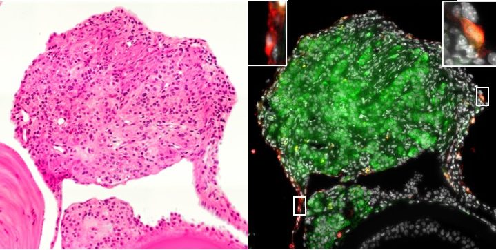
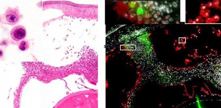
The In Vivo Modelling Core enables transplantation of primary or stem cell derived beta-like cells using encapsulated or non-encapsulated procedures, and provides a consistent approach to immune and beta-cell-based readouts. Building upon methods for anterior chamber eye transplantation the core is also developing methodology for longitudinal in vivo imaging of fluorescently labelled/reporter cells.

The images on this page is kindly provided by Dr Majid Mojibian from the Levings lab.

Images of a non-rejected human islet retrieved from an islet transplant in the anterior chamber of the eye. Left image shows a classical histology image. The right image shows the same human islet now fluorescently labelled for insulin (green) and an immune cell marker (red). Notice the difference between this non-rejected islet and the rejected islet below.
Images of a rejected human islet retrieved from an islet transplant in the anterior chamber of the eye. Left image shows a classical histology image. The right image shows the same human islet now fluorescently labelled for insulin (green) and an immune cell marker (red).